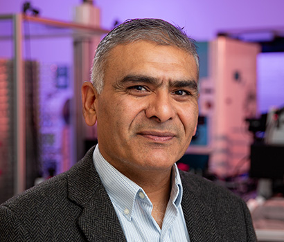
We are a team of research scientists, engineers, clinical scientists, and data scientists with deep expertise and accomplishments, working together to combine science and advanced engineering to create innovative therapies that will improve and extend life. We are endlessly curious, intellectually honest and thrive in exploring biology in new ways.
eikon therapeutics
We seek to advance breakthrough therapeutics through the purposeful integration of engineering and science.
Our Approach
Purposeful integration to create innovative therapies.
At Eikon, we discover and develop new medicines by reimagining what is possible — inventing new technologies to bring to light quantitative information about the behavior of biological systems — to identify new targets and bring novel therapies to patients.
Technology & Platform
We leverage what is available —
and invent what is not.
As experienced researchers and clinicians, we apply traditional, high-performance tools when we can. And our engineering skills allow us to build new instruments when we need them. With this approach we explore cell and molecular biology in completely new ways.
Learn More
Proprietary Hardware
Learn More
AI & Machine Learning
Learn More
Custom Reagents & Automation
Our Proprietary Technology
There is a direct link between protein motion and activity. By measuring the dynamics of protein populations, we can explore the activity of novel pharmaceuticals in great detail.
With our proprietary single-molecule tracking (SMT) platform, we can visualize hundreds of thousands of protein motion events in living cells in less than a second. By integrating AI capabilities and advanced automation together with SMT, we can inventory molecular interactions with extraordinary scale and precision.
Single Molecule Tracking (SMT) Technology
Protein activity is mediated by transient interactions between a protein molecule and other components of the cellular environment, including membranes, other proteins, DNA/RNA and small molecules.
These transient interactions define the motion profile of a protein population. By precisely measuring these motions, we describe the activities and the functions of a protein.
Pipeline
We’re delivering important new medicines for serious diseases.
Eikon has a growing therapeutic pipeline with the potential to deliver important new medicines to address serious illnesses, many with severely unmet medical needs. We have clinical programs that have already provided evidence of activity in the treatment of solid tumors.
Our research teams are rapidly advancing numerous preclinical programs across a broad range of therapeutic areas. Beyond oncology, we invest substantial effort in immunology and neuroscience.
Program
Discovery
Lead Op
IND-Enabling
Phase 1
Phase 2
TLR7/8
PARP1
PARP1 – CNSPenetrant
AR
VCP
Undisclosed
Undisclosed
AR-v7
Undisclosed
Undisclosed
Oncology
Immunology
Neuroscience
Our Leadership
Our diversity of expertise and perspectives is our strength.
At Eikon Therapeutics, we are exploring new dimensions in biology by transcending disciplines. We have assembled a team with expertise in drug discovery, clinical development, data analytics and engineering. We value a diversity of experience and perspectives and actively listen and learn from each other.
Leadership
Board
SAB
Investors


Join us on our mission
To advance breakthrough therapeutics through the purposeful integration of engineering and science
Innovate
We innovate to solve difficult problems.
Collaborate
We collaborate to build a great company.
Execute
We execute to deliver results with rigor and urgency.
Respect
We respect one another and create an inclusive culture.
Transparent
We are transparent in our interactions.
Care
We care about patients and making a difference in the world.
Start a Conversation
Join our team of renowned experts working together to achieve our mission.
Safety Reporting
To report a safety concern or adverse event (side effect) related to an Eikon Therapeutics product, please email saereporting@eikontx.com.


